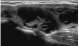
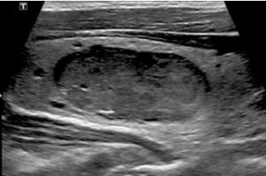
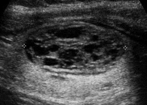
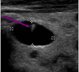
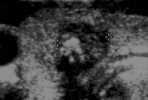
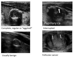
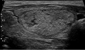
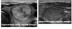
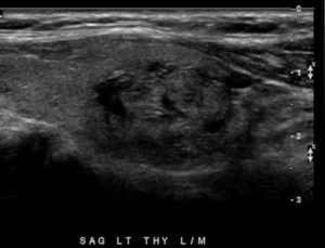
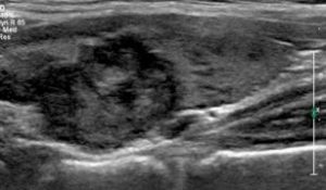
- Cystic or almost completely cystic
- Mixed cystic and solid
- Solid or almost completely solid
- Spongiform
- Large comet tail artifacts
- Macrocalcifications
- Peripheral rim calcifications
- Punctate echogenic foci
Composition
Cystic or almost completely cystic
- Cystic: Entirely fluid filled
Mixed cystic and solid
- Mixed cystic and solid: Assign points for predominant solid component.
Solid or almost completely solid
- Solid or almost entirely solid: Entirely or nearly entirely soft tissue with only a few tiny cystic spaces
Spongiform
- Spongiform: Composed predominantly (>50%) of small cystic spaces.
Echogenic Foci
Large comet tail artifacts
- Comet-tail artifacts: Deeper echoes attenuated with decreased width resulting in a triangular shape. V-shaped, >1 mm, in cystic components.
Macrocalcifications
- Macrocalcifications: Calcifications with posterior acoustic shadowing
Peripheral rim calcifications
- Peripheral calcifications: Calcifications at periphery of the nodule. Complete or incomplete along margin.
Punctate echogenic foci
- Punctate echogenic foci: < 1 mm with no posterior acoustic shadowing. May have small comet-tail artifacts.
Echogenicity
Anechoic
- Anechoic: Applies to cystic or almost completely cystic nodules.
Hyperechoic or isoechoic
- Hyperechoic: Increased echogenicity relative to the thyroid tissue
- Isoechoic: Similar echogenicity relative to the thyroid tissue
Hypoechoic
- Hypoechoic: Decreased echogenicity relative to the thyroid tissue
Very hypoechoic
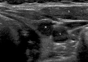
- Very hypoechoic: Decreased echogenicity relative to the adjacent neck musculature
- Very hypoechoic nodule. Margins are smooth. Note that nodule is less echogenic than adjacent strap muscles (S) and essentially isoechoic to the common carotid artery (C).
Margin
Extra-thyroidal extension
- Extrathyroidal extension: Nodule extends through the thyroid capsule. Obvious invasion = malignancy.
Ill-defined
- Ill-defined: Border difficult to discern from thyroid parenchyma
Lobulated or irregular
- Irregular margin: Spiculated, jagged or sharp angles even if only in part of the nodule
- Lobulated: Focal round soft tissue protrusions into adjacent parenchyma that may be single or multiple
Smooth
- Smooth: Uninterrupted, well-defined, curvilinear edge typically spherical or elliptical in shape
Shape
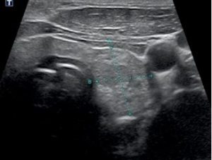
Taller-than-wide
- Taller-than-wide: Ratio of AP diameter to the horizontal diameter is > 1 when measured in the transverse plane
- Should be assessed on a transverse image with measurements parallel to sound beam for height and perpendicular to sound beam for width.
- This can usually be assessed by visual inspection.

